Stop the Cycle of Calf Pain: How to Restore Your Mobility
Have you ever felt a sudden “pop” in your calf while playing sports, or do you wake up with stiff, aching ankles every morning?
If you’re struggling with recurring calf strains or feel like your ankles just won’t bend the way they used to, you aren’t just “getting older.” There is likely a mechanical reason for your discomfort. By addressing the root cause today, you can avoid long-term damage and get back to the activities you love.
Is Your Calf Muscle Trying to Tell You Something?
Most people think a calf strain is just a one-off accident. However, if your calf pain keeps coming back, your body is signalling an underlying imbalance.
The “Sudden Snap” (Gastrocnemius Strain)
Common in tennis, running, and football, this feels like being kicked in the back of the leg.
-
The Risk: If you just “walk it off,” the muscle heals with stiff scar tissue. This makes the muscle less stretchy and much more likely to tear again.
The Deep Ache (Soleus Strain)
This is a duller, deeper pain lower down your leg. It usually hurts most when you try to push off or walk on your tiptoes.
What to do right now: If you’ve just hurt yourself, remember R.I.C.E. (Rest, Ice, Compression, Elevation). But don’t stop there—get a professional assessment to ensure you aren’t building up permanent scar tissue.
The Hidden Culprit: Ankle Equinus
Many of our patients come in for heel pain or arch aches, only to find out the real problem is Ankle Equinus.
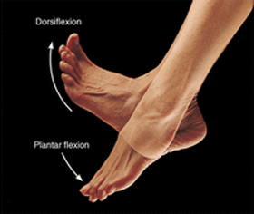
In plain English: This just means your ankle doesn’t bend upward enough (less than 10 degrees). Because your ankle is “locked,” your body has to cheat to help you walk. This “cheating” causes a chain reaction of pain:
-
The “Heel Lift”: You lift your heel too early, putting massive pressure on the ball of your foot.
-
The “Flatfoot”: Your arch collapses to try and find the flexibility your ankle is missing.
-
The “Toe Walk”: You find yourself walking on your toes or swinging your hips unnaturally.
Why is my ankle so stiff?
It’s usually not your fault! It can be caused by:
-
Tight Achilles Tendons: Often something you’re born with or inherited.
-
Lifestyle: Frequent use of high heels or previous injuries in a cast.
-
Bone Blocks: Small fragments of bone from an old injury physically stopping the joint from moving.
How We Get You Moving Again
You don’t have to live with “tight” legs. We use simple, non-invasive methods to unlock your mobility and take the pressure off your joints:
-
Custom Orthotics: Specially made inserts that re-balance your weight so your muscles don’t have to work overtime.
-
Night Splints: A gentle way to stretch your calves while you sleep—no effort required!
-
Heel Lifts: A simple “boost” inside your shoe to take the immediate golden-thread tension off your Achilles.
-
Smart Stretching: Targeted exercises that actually work for your specific body type.
Take the First Step to Pain-Free Living
Ignoring a stiff ankle or a tight calf today often leads to Plantar Fasciitis, Shin Splints, or Back Pain tomorrow. The sooner you fix the mechanics, the faster you’ll be back on the court, the track, or just enjoying a walk without पहुँचा discomfort.
Ready to find out why your feet are aching?
[Book Your 30-Minute Foot & Ankle Assessment] — Let’s find the root cause together.